Patella stabilisation surgery aims to prevent repeated dislocation or instability of the kneecap (patella). When the patella slips out of place, it can cause pain, swelling, and damage to the joint surface. In many cases, instability occurs because the medial patellofemoral ligament (MPFL)—the main soft-tissue restraint that stops the kneecap from moving outwards—has been stretched or torn during a dislocation.
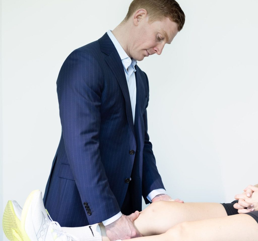
While there are several surgical techniques to stabilise the patella, the most common for patients with recurrent instability is MPFL reconstruction. This procedure restores the ligament’s function, helping keep the kneecap centred in the groove at the end of the thigh bone, especially during early knee bending.
Occasionally, in the presence of abnormal anatomy, additional procedures may be recommended. These can include tibial tubercle osteotomy, trochleoplasty, or femoral or tibial derotation osteotomies. Dr Free is experienced in all forms of patella stabilisation surgery and will discuss the most appropriate option for your individual needs.
MPFL reconstruction replaces the damaged ligament with a graft – commonly taken from one of your own hamstring tendons or, in some cases, from a donor. The graft is fixed to the inside of the kneecap and thigh bone using small anchors or screws. This new ligament acts as a check-rein to stop the patella from sliding sideways.
The surgery is performed through small incisions. Dr Free also performs a knee arthroscopy at the start of the procedure to assess for any additional injuries and treat any cartilage damage.
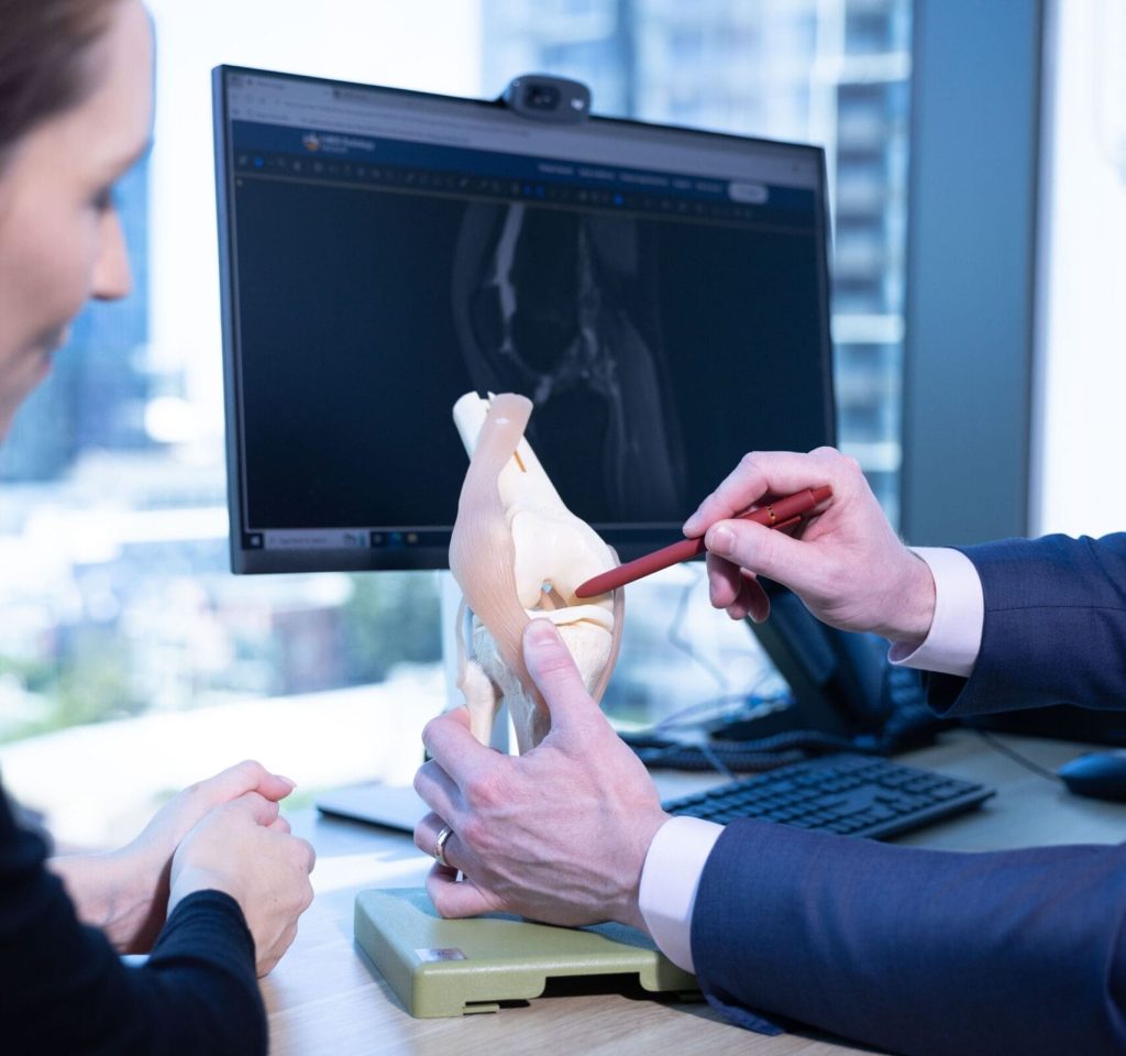
A tibial tubercle osteotomy is a surgical procedure that repositions the bony attachment of the patellar tendon on the tibia (shinbone). By shifting this attachment point, the alignment of the kneecap can be improved, reducing the tendency for it to dislocate or track abnormally. The surgery involves making a controlled cut in the bone, moving the tibial tubercle to the desired position, and securing it with screws. This procedure is often performed alongside MPFL reconstruction when patella instability is associated with abnormal bony alignment.
A trochleoplasty is a specialised surgical procedure designed to reshape the trochlear groove — the groove at the end of the thigh bone (femur) in which the kneecap moves. In some people, the groove is abnormally shallow or even convex, which makes the patella more prone to dislocation. Trochleoplasty deepens and reshapes the groove, improving patella tracking and stability. This is typically considered in patients with significant trochlear dysplasia and is often combined with MPFL reconstruction or other stabilisation procedures.
Please refer to the Pre-operative information page for guidance on preparing for your procedure.
Most patients stay in hospital overnight following surgery.
Most patients can walk with the aid of crutches immediately after surgery. A brace may be used for up to 6 weeks post-surgery. Swelling control and regaining range of motion are critical in the first few weeks. A full return to sport depends on your progress with rehabilitation and choice of sport, however for MPFL reconstruction this is often around 4-6 months after surgery.
See my MPFL reconstruction rehabilitation protocol for full details.
Dr Free has extensive experience in diagnosing and treating patella instability, tailoring personalised treatment plans that address each patient’s unique anatomy and lifestyle. He carefully evaluates all surgical options to recommend the most appropriate procedure for lasting knee stability.
Around 95% of patients experience no further dislocations, with improved confidence and function in the knee.
Without surgery, the risk of repeated dislocations remains high, which can lead to cartilage damage, early arthritis, and reduced knee function over time.
Yes — rehabilitation is crucial. Physio begins within the 1-2 weeks and focuses on restoring motion, building strength, and gradually returning you to sport.
Most patients will use crutches for 2-4 weeks after surgery. As a general rule, crutches should be used until you have regained full range of motion and are walking normally without a limp.
You should avoid driving until you are off strong pain medications, walking without crutches, and have good knee range of motion and muscle control. This is usually around 6 weeks but may be sooner for left knee surgery with an automatic car. Always check with Dr Free before resuming driving.
The wound should be kept dry until your review with Dr Free at 2 weeks post-surgery. Following this, you will be able to shower with no dressing on. Swimming pools or hot tubs should be avoided for 3-4 weeks to minimise the risk of infection.
Return to sport is typically around 4-6 months after surgery, depending on your recovery with rehabilitation, sport type, and whether additional procedures were required. Dr Free and your physiotherapist will assist you in planning your return to sport.
As with any surgery, there are risks including infection, stiffness, or over-tightening of the graft. Other risks include skin numbness around incisions, kneecap fracture, or recurrent instability. There is also a small risk of complications related to any additional procedures, such as bone healing issues after a tibial tubercle osteotomy. These risks will be discussed in detail during your consultation.
Dr Matthew Free offers expert, personalised solutions for every step of the journey.